 section 1
 section 2
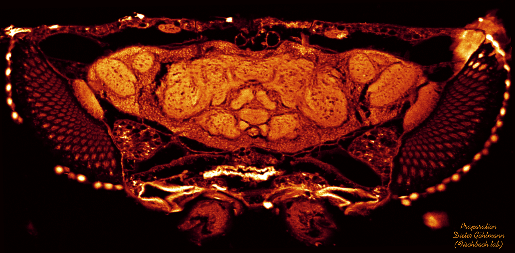
 section 3
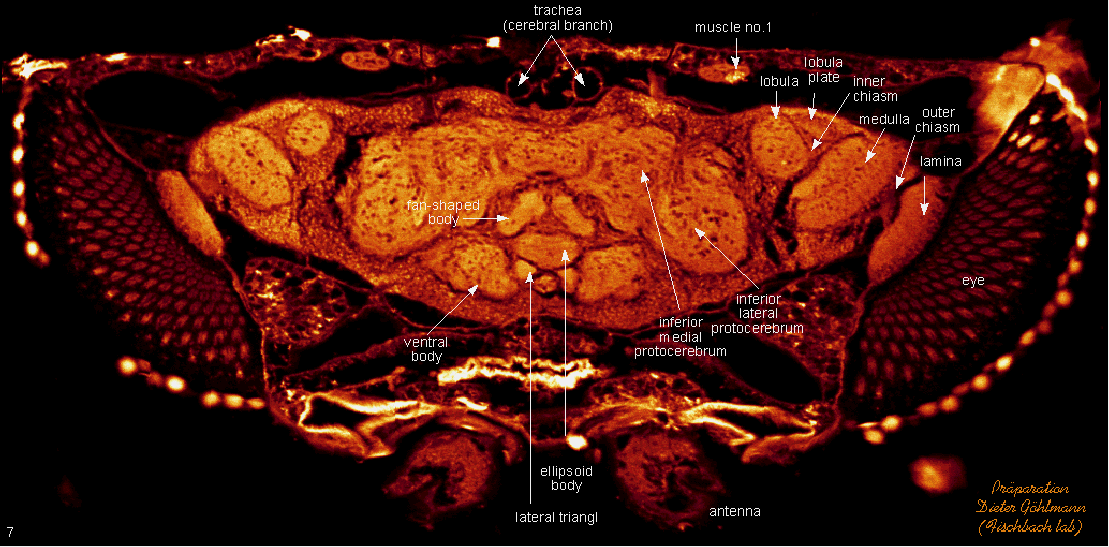
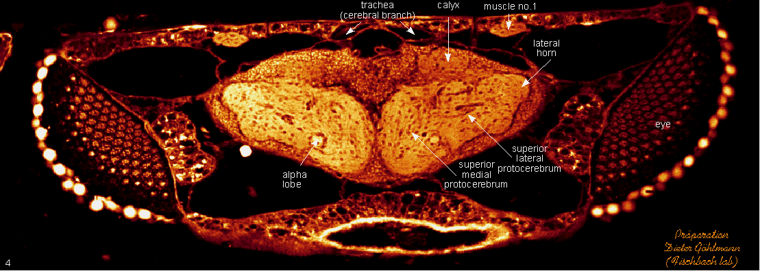
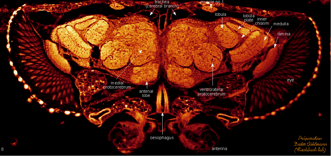
 section 4
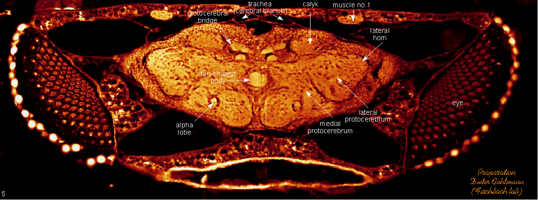
 section 5
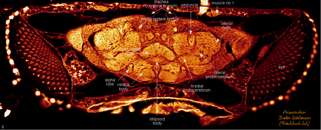
 section 6
 section 7
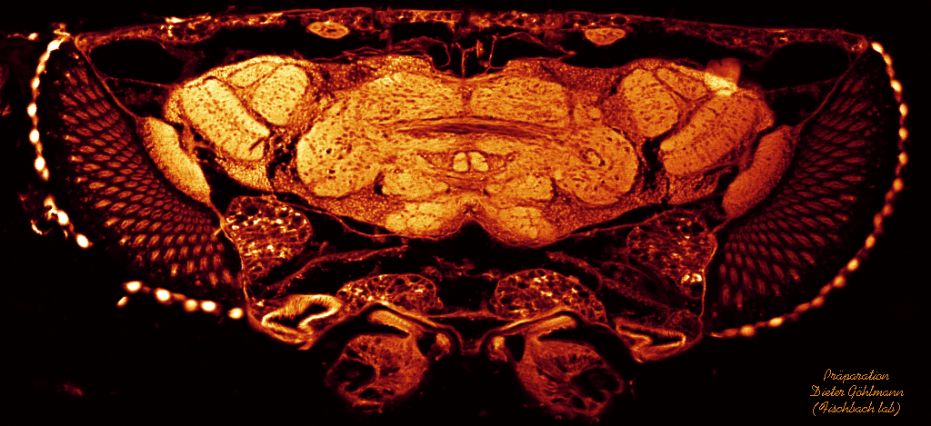
 section 8
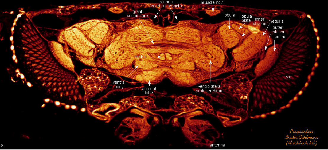
 section 9
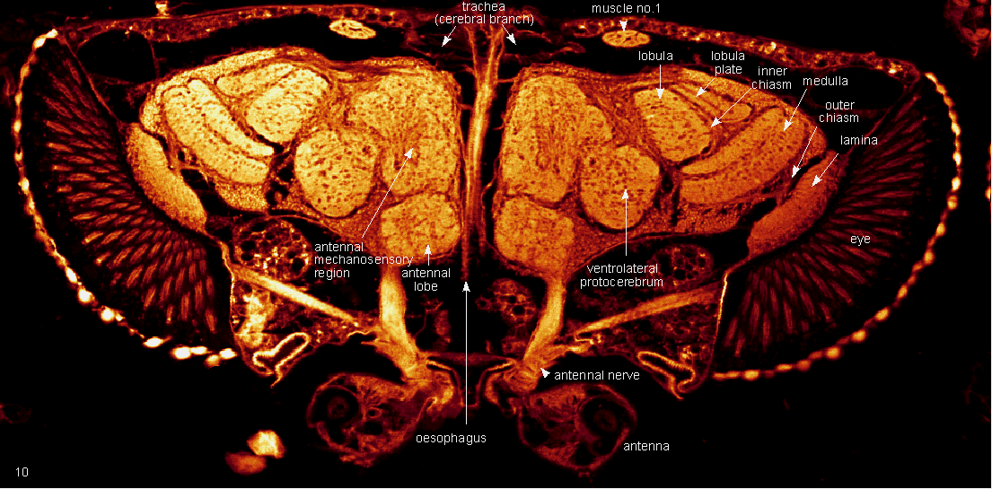
 section 10
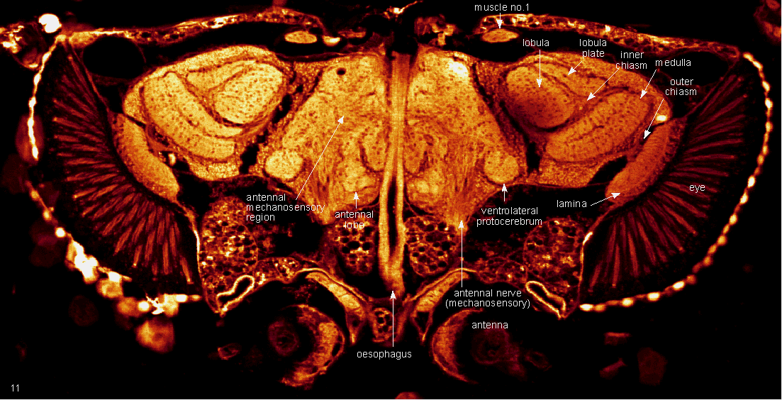
 section 11
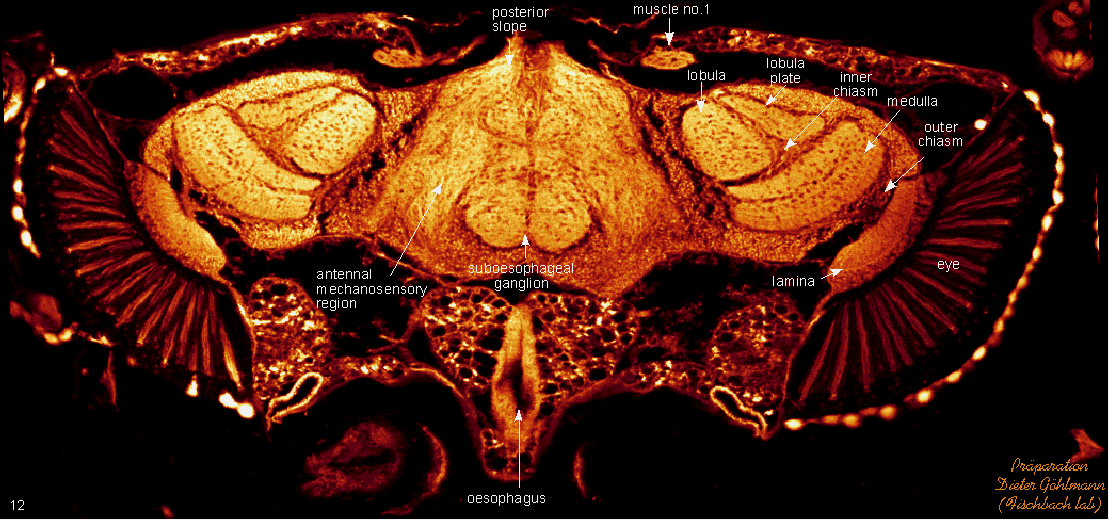
 section 12
 section 13
 section 14
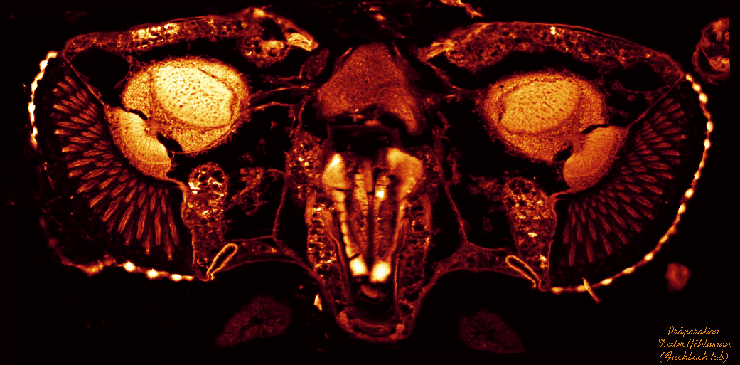
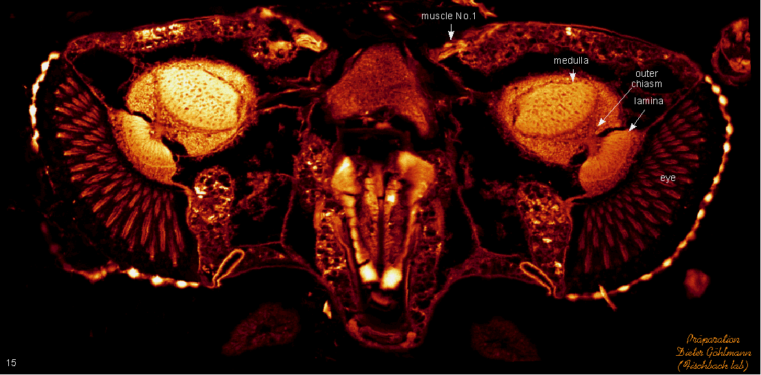
 section 15
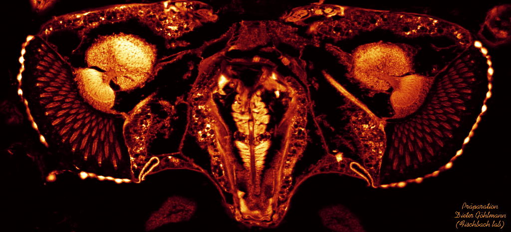
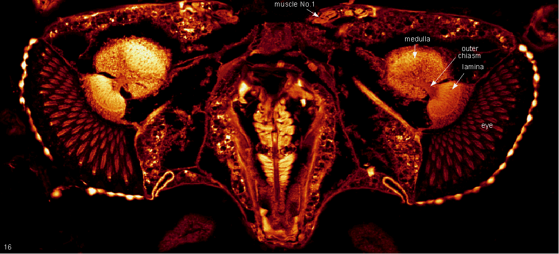
 section 16
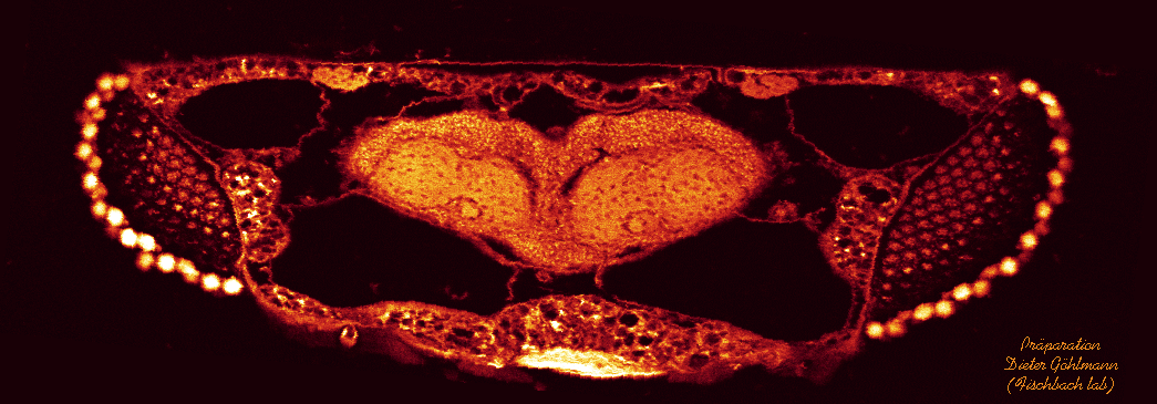
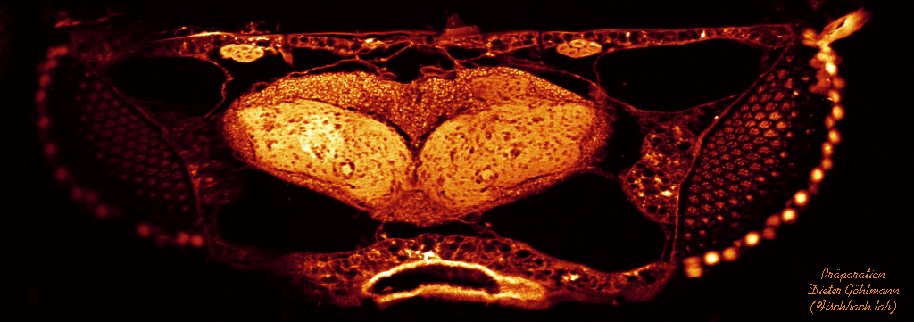
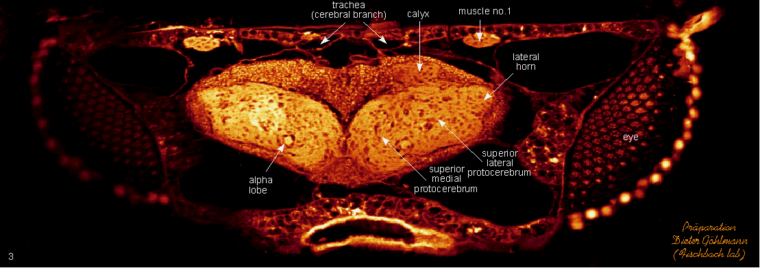
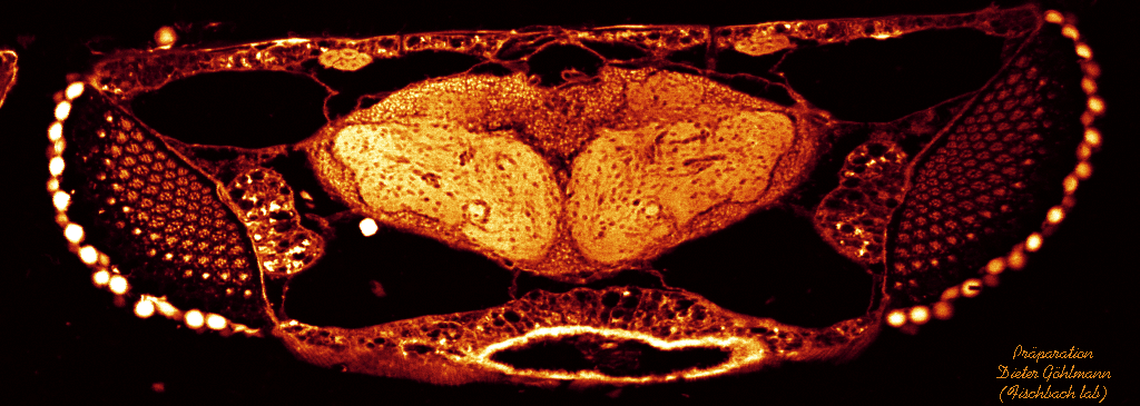
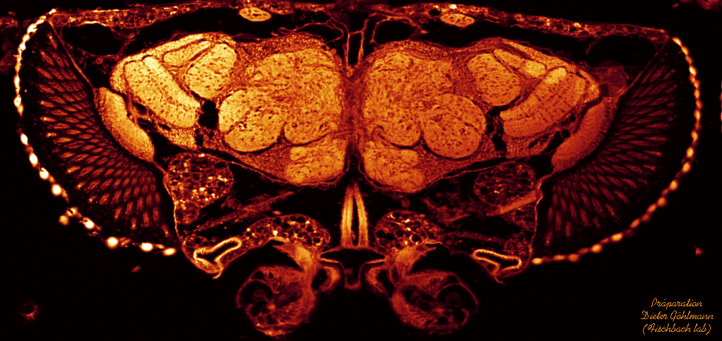
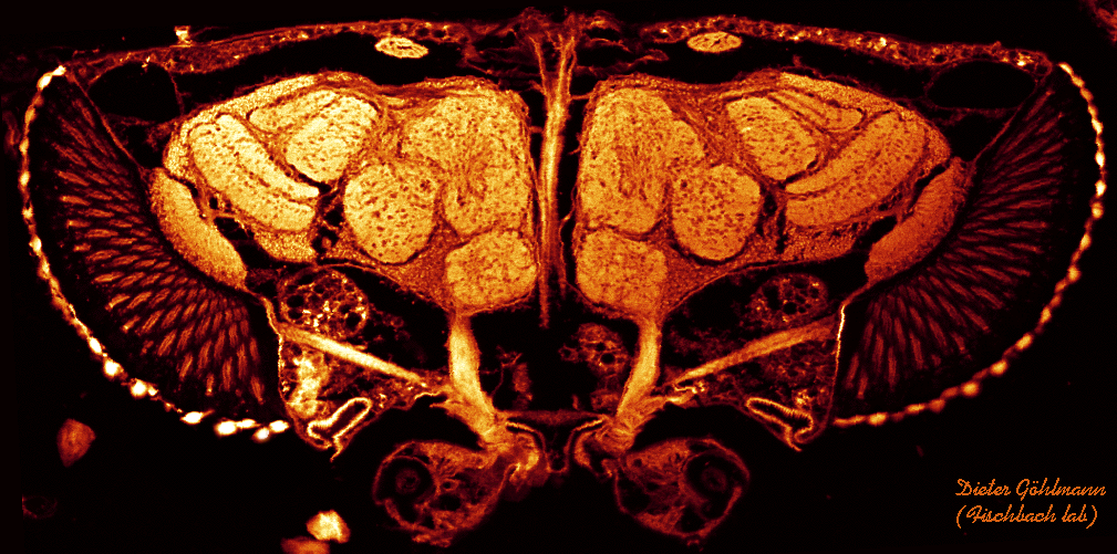
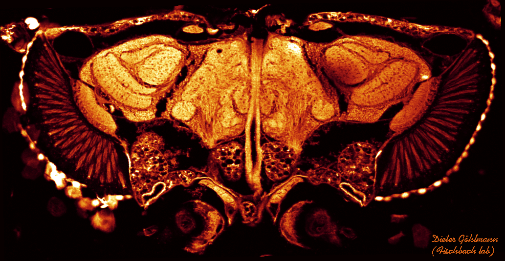
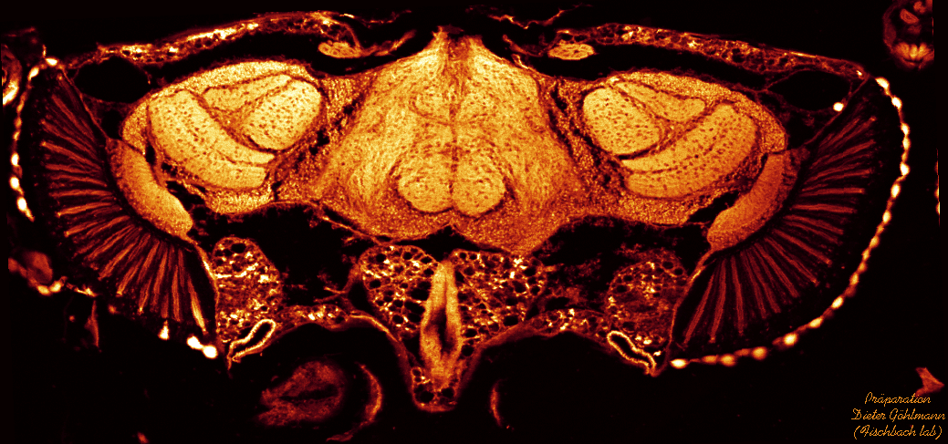
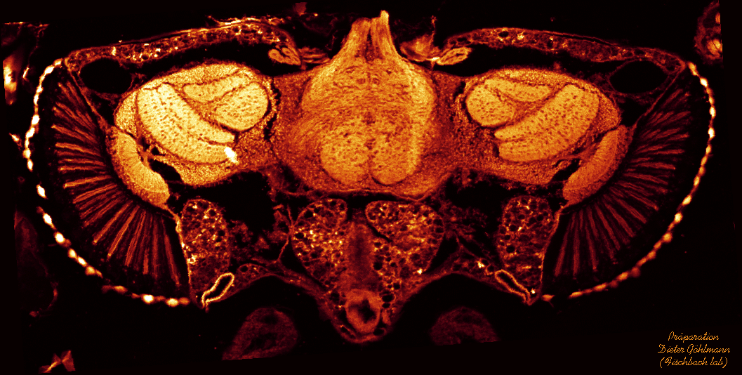
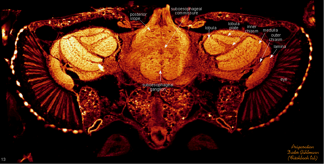
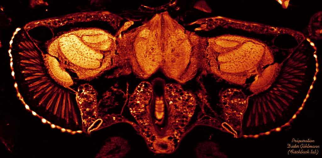
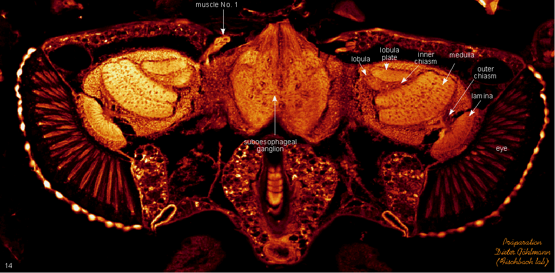
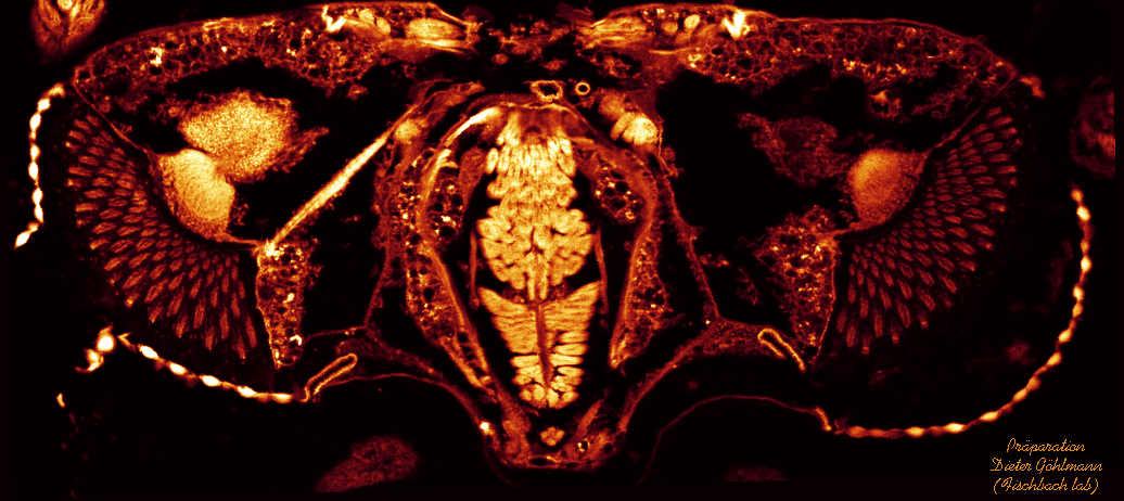
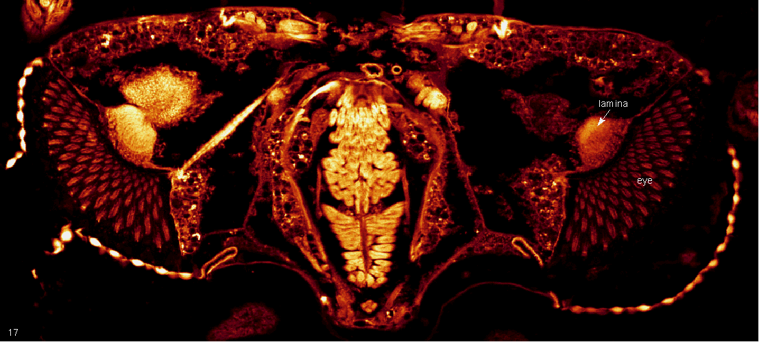
Sixteen 7 µm horizontal paraffin sections of male flies. Specimen were prepared following the collar method. Autofluorescent sections were viewed in the confocal microscope and scanned in 1024 x 1024 mode. Sections can, of course, also be viewed under a normal fluorescent miscroscope. In this case, they have a greenish colour.
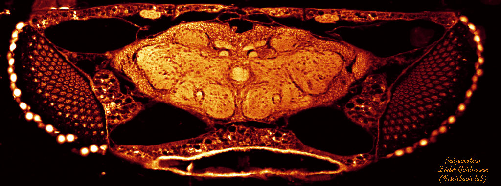
Click on thumbnails to get clean section, click on "section x" for labeled section.
Be patient while downloading, it will pay. If downloading takes too long, you can open a second window of your browser and continue web surfing there.
section 1
section 2
section 3
section 4
section 5
section 6
section 7
section 8
section 9
section 10
section 11
section 12
section 13
section 14
section 15
section 16