Lamina-medulla connections in sol mutants
Andreas Dittrich and Karl-Friedrich Fischbach, unpublished.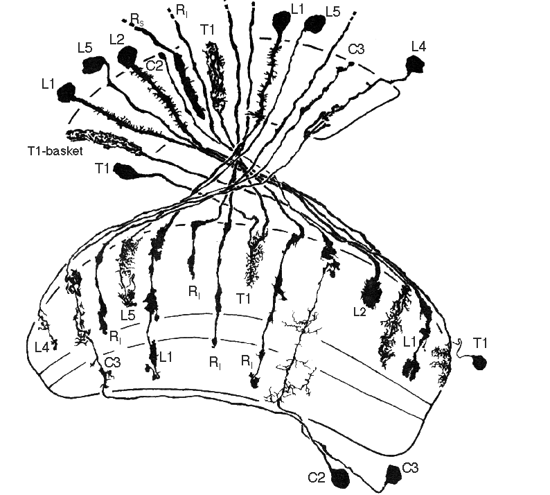
Click for full resolution (85K)
Camera lucida drawings of columnar neurons connecting lamina and medulla in the small optic lobes mutant allele KS58. In comparison with the wild type situation it is clear that most of the neurons known are present in sol . While their neuronal shapes are pretty normal at the level of the lamina, their arborizations at the level of the medulla tend to be less stratified (one of the reasons why L3 might be difficult to find). Long retinal fibers (Rl) and L1-terminals may extend into the proximal medulla.
L1-L5 = lamina monopolar neurons 1 - 5 with their cell bodies in the lamina cortex
T1 = T1 neuron with its cell body in the medulla cortex and arborizations in medulla and lamina (forming so-called baskets in the lamina).
C2,3 = Centrifugal cells 2 and 3 with their cell bodies distal to the lobula plate cortex, dendritic arborizations in the medulla and club-like terminals in the lamina.
Rs = terminal of short retinula cell (of the R1-6 type).
Interpretation: These data agree with the volumetric measurements showing that the volume of the lamina is about normal in sol . The altered (enlarged) arborizations in the medulla of the neurons shown presumably are their response to the absence of about 50% of all columnar medulla neurons. Genetic mosaic analysis has shown that the genotype of the medulla cortex is responsible for the mutant phenotype (small optic lobes resulting from cell degeneration in the pupal stage; Fischbach K.-F. and Technau, G. (1984)).